


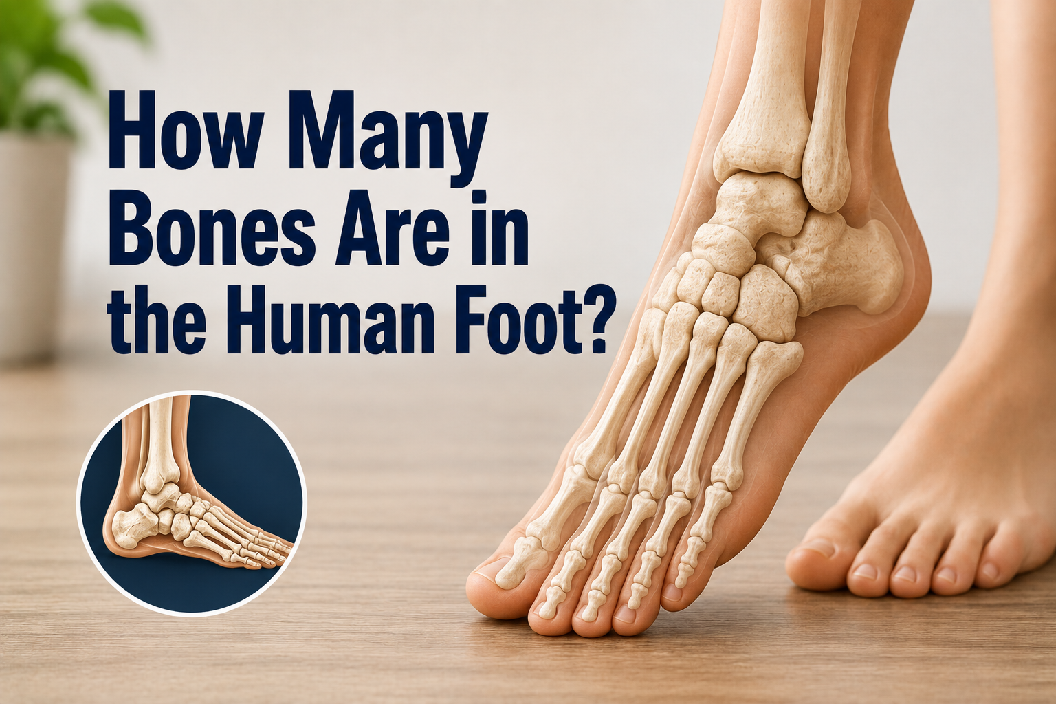
The human foot contains 26 individual bones, along with two small sesamoid bones tucked beneath the big toe joint. Together, these bones make up close to a quarter of all the bones found in the entire human body, which shows just how complex this small structure really is.
When people ask how many bones human foot has, the short answer is 26 per foot, or 52 bones across both feet combined. These bones are grouped into three main regions: the tarsals near the ankle, the metatarsals in the middle of the foot, and the phalanges that form the toes.
This bone arrangement is what allows the foot to absorb shock, support body weight, and adapt to uneven ground while walking or running. The rest of this guide breaks down each group in detail so you understand exactly where every bone sits and what job it performs.
The Three Main Regions of the Foot
Every foot bone belongs to one of three connected regions, and each region has its own shape and purpose. The sections below explain the tarsals, metatarsals, and phalanges individually so the full structure is easy to follow.
Tarsals
The tarsal bones sit at the back of the foot, closest to the ankle, and there are seven of them in total. They form a sturdy cluster that connects the leg to the rest of the foot and carries most of the body’s weight when standing. Because of their thickness and dense packing, this region is the most stable part of the entire foot.
Metatarsals
The metatarsals are five long, narrow bones located in the middle section of the foot. Each one runs from the tarsal bones toward a specific toe, acting like a bridge between the back of the foot and the toes. These bones flex slightly with every step, which helps push the body forward while walking or running.
Phalanges
The phalanges are the smallest bones in the foot, and there are fourteen of them making up the toes. Each toe except the big toe has three phalanges, while the big toe only has two. These small bones give the toes flexibility and help maintain balance during standing and movement.
Tarsal Bones Explained
The seven tarsal bones each have a distinct name, shape, and role within the ankle and hindfoot region. Understanding what each one does makes it easier to see why this area is so important for stability.
Talus
The talus sits directly below the shinbone and forms the main connection point between the leg and the foot. It transfers the body’s weight downward into the heel and the rest of the foot with every step taken. Unlike most bones, no muscles attach directly to the talus, which makes it dependent on surrounding ligaments for support.
Calcaneus
The calcaneus, commonly known as the heel bone, is the largest bone in the entire foot. It absorbs the initial impact whenever the heel strikes the ground while walking, running, or jumping. Because it bears so much repeated stress, the calcaneus is also one of the most common sites for foot fractures.
Navicular, Cuboid, and Three Cuneiforms
These five remaining tarsal bones sit in a row between the talus and calcaneus at the back and the metatarsals at the front. The navicular and cuboid bones act as connectors, while the three cuneiform bones help form the arch of the foot. Together, this group gives the midfoot its shape and allows small, controlled movements needed for balance.
| Bone Name | Location in Foot | Primary Function |
| Talus | Top of hindfoot, below shinbone | Transfers body weight from leg to foot |
| Calcaneus | Back of hindfoot | Absorbs heel-strike impact |
| Navicular | Midfoot, inner side | Connects talus to cuneiform bones |
| Cuboid | Midfoot, outer side | Supports outer arch structure |
| Medial, Intermediate, Lateral Cuneiforms | Midfoot, front row | Form the foot’s arch and connect to metatarsals |
Metatarsal Bones Explained
The five metatarsal bones are numbered and positioned in a way that mirrors the layout of the toes above them. Below, the numbering system and common injury patterns are explained separately for clarity.
Numbering System
Metatarsal bones are labeled first through fifth, starting from the big toe side and moving toward the little toe side of the foot. The first metatarsal, connected to the big toe, is the shortest and thickest of the group since it carries the most weight. The fifth metatarsal, on the outer edge of the foot, is thinner and more prone to injury during sideways movements.
Common Injuries
Stress fractures are frequent in the metatarsals, particularly among runners and athletes who place repeated pressure on the midfoot. The fifth metatarsal is especially vulnerable to a specific break known as a Jones fracture, which often occurs from twisting the ankle. These injuries usually cause swelling and pain that worsens with walking, so early diagnosis helps prevent long-term complications.
Phalanges — The Toe Bones
The toes contain fourteen small bones total, but they are not distributed evenly across all five toes. The next two sections explain why the big toe differs structurally from the rest.
Big Toe vs. Other Toes
The big toe, also called the hallux, has only two phalanges instead of three like the other toes. This shorter structure makes the big toe stronger and better suited for pushing off the ground during walking. The remaining four toes each contain a proximal, middle, and distal phalanx, giving them more flexibility but slightly less strength.
Why the Big Toe Differs Structurally
The big toe evolved with fewer bones because its main role is weight-bearing and propulsion rather than fine movement. This design allows it to handle a significant portion of the force generated during each step, especially when pushing off to walk or run. The smaller toes, by contrast, contribute more to balance and grip on uneven surfaces.
Sesamoid Bones — The “Hidden” Bones
Beneath the joint of the big toe sit two small, pea-shaped bones known as sesamoid bones, which are not always included in the standard count of 26.
These bones are embedded within a tendon rather than connected directly to other bones, which is what makes them unique compared to the rest of the foot’s skeleton.
Their main job is to reduce friction and protect the tendon as it moves during walking, running, and jumping. Because of their position, sesamoid bones can become irritated or fractured through repetitive stress, a condition often seen in runners and dancers.
Foot Bones vs. Other Joints and Ligaments
Bones give the foot its basic shape, but movement itself depends on joints and ligaments working alongside those bones. Joints are the points where two or more bones meet, while ligaments are the fibrous tissues that hold those joints together and limit excessive motion. The subtalar joint, located between the talus and calcaneus, allows side-to-side rocking of the foot.
The ankle joint connects the leg bones to the talus and permits up-and-down movement, while the midtarsal joint allows twisting motion through the middle of the foot. Without healthy ligaments supporting these joints, even a perfectly intact set of 26 bones would not be able to move properly.
Common Foot Bone Conditions
Foot bones are subject to a range of conditions caused by repetitive stress, structural imbalance, or sudden injury. The three most frequent categories are explained individually below.
Fractures
Fractures in the foot can range from minor stress fractures caused by overuse to more severe breaks from a direct impact or fall. The metatarsals and calcaneus are the most commonly fractured bones due to the amount of pressure they absorb daily. Symptoms typically include sharp pain, swelling, and difficulty putting weight on the affected foot.
Bunions and Bone Spurs
A bunion forms when the joint at the base of the big toe becomes misaligned, causing a bony bump to develop on the side of the foot. Bone spurs, on the other hand, are extra bone growths that often develop around joints due to long-term stress or arthritis. Both conditions can cause discomfort while walking and may worsen if left untreated over time.
Flat Feet and High Arches
Flat feet occur when the arch formed by the tarsal and metatarsal bones does not develop or maintain its normal curve. High arches are the opposite condition, where the arch sits unusually elevated compared to a typical foot structure. Both variations can affect how weight is distributed across the foot bones during standing and walking.
When to See a Doctor
Most everyday foot discomfort resolves with rest, but certain symptoms point to a more serious bone-related problem. Sudden swelling that does not go down, visible deformity, or an inability to bear any weight on the foot are signs that a fracture or dislocation may be present.
Persistent pain that lasts more than a few days, especially after an injury, twist, or fall, should also be checked by a medical professional. Getting an evaluation early, often through an X-ray, helps prevent a minor injury from turning into a long-term mobility problem.
Frequently Asked Questions
Do babies have the same number of foot bones as adults?
No, babies are actually born with more foot bones than adults because several of these bones start out as separate pieces of cartilage. Over time, some of these pieces fuse together as the child grows, eventually forming the standard 26-bone structure seen in adults.
Why do feet have so many bones compared to other body parts?
Feet need this many bones because they perform two very different jobs at once: supporting body weight and adapting to uneven surfaces. A smaller number of bones would make the foot far more rigid, reducing its ability to absorb shock and adjust while walking or running.
Can you lose a foot bone and still walk normally?
In some cases, yes, particularly with smaller bones like certain phalanges, since surrounding bones and tissue can partially compensate. However, walking patterns are often affected to some degree, and the extent depends on which bone was lost and how the surrounding structure adapts.
Are all 26 bones necessary for balance?
While every bone contributes something to overall foot function, the tarsal bones play the largest role in maintaining balance due to their position near the ankle. The metatarsals and phalanges assist with balance mainly during movement, such as walking, running, or standing on uneven ground.
Conclusion
The human foot’s 26 bones work together as a single, coordinated system rather than as isolated parts. From the sturdy tarsals near the ankle to the flexible phalanges at the toes, each bone group plays a specific role in supporting weight, absorbing impact, and enabling smooth movement.
Compared to generic anatomy explanations found elsewhere, this breakdown focuses on how each bone group connects to real, everyday foot function rather than just listing names. Understanding this structure makes it easier to recognize when foot pain might point to something more serious than everyday fatigue.
RELATED ARTICLES
Itchy Inner Thighs: Causes, Symptoms, Treatments, Home Remedies, and Prevention
How to Unlock a Locked Knee: Causes, Immediate Steps & When to See a Doctor
Can Bad Posture Cause Chest Pain? Causes, Symptoms & Relief
How to Cure Burning Watery Eyes: Causes, Remedies & When to See a Doctor